Bone scan is one of the nuclear scanning procedures used to detect various bone deformities or conditions. This procedure is highly sensitive to osteoblast activities that cause abnormal bone overgrowth in various parts of the body. Other radiologic diagnostics include Magnetic Radioactive Imaging (MRI), Positron Emission Tomography (PET) and X-ray. In X-ray, a radiation is sent through the body to project the structure of the bones. In this procedure, the radiation is emitted from the inside. It costs less than other radiologic procedure yet highly effective in determining bone metabolism abnormalities.
When is the Bone Scan Conducted?
There are many reasons why a bone scan is required. It is used when suspecting bone trauma after serious accidents wherein there is stress and fracture on the bone. It can also effectively measure the severity and age of fractures. It is also utilized when X-ray is unable to localize abnormal bone pain. Bone scan is used as well to detect abnormalities in bone growth and structure as such in Paget’s disease and arthritis. These diseases can be debilitating and early diagnosis can be very beneficial to the bearer.
Bone infection and inflammation such as on the joints or shaft can also be diagnosed through this procedure. Bone infection diagnosed with this procedure includes osteomyelitis. This is also done to detect abnormal blood supply in different parts of the bones. Lastly, this is used to detect bone cancer or any cancer that has metastasized to the bones.
Preparation and Precautions before Bone Scan
Be sure to inform your doctor, before the bone scan procedure, if you are pregnant or breastfeeding. Pregnant women can postpone the test since this could affect the development of the fetus due to some radioactive activity inside the body. For those who are breastfeeding, it is best to avoid doing so until 4 days after the test to allow all the radioactive dye to be flushed out of the body.
Be sure that there is no intake of Bismuth or Barium containing medications up to 4 days before the test as this could significantly alter bone scan results. This could generate false-negative results that may affect treatment and medication required.
It is also advisable to limit fluids hours before the dye is injected and increase intake 1-2 hours after. This is to flush the extra dye that was not absorbed by the bones. It is also important that the bladder is not full during the scan to have the fullest visualization on the pelvis area. Although it only emits trivial amount of radioactivity, it is best to ensure proper hygiene and hand washing after voiding.
Bone Scan Procedure
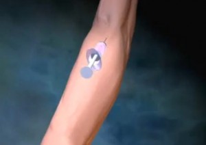
The bone scan is basically divided into 2 parts. The first part would be the injection of the medium dye through the vein in the arm. The second is the scan itself which is done 2 to 4 hours after. In between the waiting period you can leave the radiologic area without causing any harm to anyone.
Bone scan procedure is able to project the bones through injection of a radioactive dye called tracers. Technetium 99 is the primary choice as it is a radionuclide that emits gamma rays. These emitted gamma rays can easily be captured by a gamma sensitive camera that takes successive images in various areas of the body. This allows proper visualization of the bone function and activity on each area taken.
As the dye is injected, it travels to the bones and different organs through the bloodstream. This is absorbed and settles in the bone at different patterns and concentrations. This is the basis of determining which part is healthy and which is abnormal. Those that are not absorbed are immediately flushed out of the body via urine. This is why it is important to increase fluid intake after the dye is injected.
The scan can last to an hour or less depending on how wide the involved area is. It is quite uncomfortable during the scan as you will have to stay still throughout the procedure. You might want to request for a comfortable pillow where you can rest your head on. Usually, the images are taken 4 hours after the dye is injected. Although in some occasions a three-phase scan is done. That is consecutive images taken at different points in the dye absorption. First, as the dye is injected. Second, right after it is absorbed. Lastly, 3 to 4 hours later.
Side Effects of Bone Scan
Bone scan is relatively safe for anyone to undergo. There are only few side effects associated with the procedure. Although localized pain and redness in the injection site can occur, bleeding and infection are remarkably rare. A few may experience allergies, rashes and even anaphylaxis, however, these can easily be addressed.
Many have second thoughts in doing so because of the radioactive dye injected in the body. This dye actually exposes the body to the most minimal radioactive activity. No healthy cells are harmed throughout the whole procedure.
Also when the dye is injected in the body it does not stay radioactive for long. It would take a full 2 to 3 days before radioactivity is completely eliminated in the body. There are no other side effects from the procedure and there is no special care needed after.
Interpretation of the Bone Scan Results
The photos taken from the scan is compiled and analyzed as to where the die is highly evident and where it is barely noticeable. A healthy bone scan would show an evenly distributed concentration of the medium dye all throughout the body.
Abnormal results are categorized as “cold spots” or “hot spots”. Cold spots are those areas that dye is less absorbed than other area. This could indicate that the blood supply in the area is insufficient. It could also suggest abnormal bone growth in the area. Hot spots are those areas that the dye has a noticeably high concentration than others. This is indicative of tumor, infection or fracture on the site.
As said, bone scan effectively establishes the site for abnormal bone metabolism, however, it would not be able to provide the exact cause for such. It is highly recommended to conduct a thorough health history on every patient in order to ascertain the proper diagnosis and treatment.
